Posts

“The Quarterly Journal of Veterinary Science in India and Army Animal Management” Vol 1 Issue 2 – January 1883

Terms of Use
This material has been identified as being free of known restrictions under copyright law, including all related and neighbouring rights, and is being made available under the Creative Commons, Public Domain Mark.

‘The Veterinarian’ Vol 56 Issue 2 – February 1883

Terms of Use
This material has been identified as being free of known restrictions under copyright law, including all related and neighbouring rights, and is being made available under the Creative Commons, Public Domain Mark.

‘The Veterinarian’ Vol 56 Issue 3 – March 1883

Terms of Use
This material has been identified as being free of known restrictions under copyright law, including all related and neighbouring rights, and is being made available under the Creative Commons, Public Domain Mark.

‘The Veterinarian’ Vol 56 Issue 4 – April 1883

Terms of Use
This material has been identified as being free of known restrictions under copyright law, including all related and neighbouring rights, and is being made available under the Creative Commons, Public Domain Mark.

“The Quarterly Journal of Veterinary Science in India and Army Animal Management” Vol 1 Issue 4 – July 1883

Terms of Use
This material has been identified as being free of known restrictions under copyright law, including all related and neighbouring rights, and is being made available under the Creative Commons, Public Domain Mark.

‘The Veterinarian’ Vol 56 Issue 9 – September 1883

Terms of Use
This material has been identified as being free of known restrictions under copyright law, including all related and neighbouring rights, and is being made available under the Creative Commons, Public Domain Mark.

‘The Veterinarian’ Vol 57 Issue 7 – July 1884

Terms of Use
This material has been identified as being free of known restrictions under copyright law, including all related and neighbouring rights, and is being made available under the Creative Commons, Public Domain Mark.

FS/2/2/2/1/9 – Selected papers from veterinary case notes relating to the Foot

Terms of Use
This material is Crown copyright, and contains public sector information licensed under the Open Government License v.3.0.

[FS/2/2/2/1/9]
[[1]]

1884 Case 83, No 45.

June 10th

Laminitis in all four feet principally the fore ones due to being overworked on first journey the Battery.

Treatment. mash diet apply wet swabs to coronet.

Stand in tank 4 hours daily

[June] 22nd

Better. repeat treatment.

July 14th

I can see no signs of Laminitis about the feet at the present time. I suspect the case must have come on more of congestive than inflammatory. Going fairly sound & sent to duty to see whether any thing develops itself.

Discharged, Relieved.

Case 117, No 45

21st July

Laminitis Chronic. This horse (see case above) went to duty and after one mornings work came back intolerably lame with every symptom of laminitis. There is not much heat in the foot & no constitutional disturbance. I therefore conclude it is more congestive than inflammatory & more chronic than acute.

Stand in foot bath.

23rd

The casting committee assembled this day & this animal was brought forward by the O.C. I gave it as my opinion that the lameness would be recurrent & that from the short time I had known the animal it was impossible for me to say that the case was perfectly incurable. Nevertheless, I believe it to be so & the animal an undesirable one to retain for further treatment. There is no alteration in the shape of the feet, no rings round the wall, but the action is most indicative of the disease.

P.T.O

[[2]]

1884

July 24th

This morning the patient was brought out again for my inspection & was found so lame that he could only just progress on the back of the heels with his fore legs right out in front of him, his hind under the body.

Cold water applications & exercise.

Augt 1st

This morning is worse than ever & the animal having been cast, I discharge him off the books.

Discharged, incurable.

Autopsy. I only found in two places, not more than one inch wide any sign of discolouration of the sensitive laminae, they being here scarlet & at their attachment nearly black. The discoloured laminae were not enlarged but were flabby & portions of them remained adherent to the horny wall where this was removed. On examining the navicular bursa of one limb it was found to contain very little synovia. The perforans tendon has eroded as if it has been scraped with a knife. The navicular bone had brown patches on its fibro cartilage on either side of the central ridge. There was no sign of ulceration, but the cartilage was speckled with calcareous deposits. The opposite navicular bursa contained no synovia but was moist, the tendon eroded & discoloured, the fibro-cartilage covering the bone was brown along its lower half & speckled with calcareous deposits: at two small points blood could be seen when the bone was bare.

Remarks. This is a very exceptional case, I believe the whole lameness was due to the diseased navicular bursa, & that the laminitis appearance was post-mortem; yet, during life no one could have seen this patient but would have said that the gait was truly indicative of acute laminitis.

Remarked during life that the feet presented no appearance of laminitis, & as will be seen from my remarks I doubted the diagnosis of my predecessor; yet when the animal came under my observation lame, I felt no doubt in my mind of the correctness of my predecessors views, & diagnosed the case, even in the absence of heat & alteration in the feet as laminitis chronic.

M/2 R.A. M 45 Sex G Age 6

Laminitis

[Transcription by Claudia Watts, KCL History, April 2019]

[FS/2/2/2/1/9]
[[1]]

Veterinary Regulations

Death Report

12th Regiment Lancers

Station: Bangalore

Date: 7th July 1883

Troop or Battery: F

Number of Horses: 42

Sex: G

Age: 15

Disease: Laminitis

Date of Admission: 1 Feby

Date of Death: 7 Feby

Record of the Case

Copies from the Record Case Book

Feby 1st

Laminitis. This is another old horse whose life has been worked out of him at the Camp of Exercise, he is off feed, dull, conjunctive dirty red.  Pulse can only be taken with difficulty owing to his being so vicious, the Temperature also cannot be taken on that account. The case is one of extreme exhaustion caused by the killing work they have just had. He won’t feed & nothing can be given him on account of his vice.

[Feby] 2

No change, stiff all over & disinclined to move. Refuses everything.

[Feby] 3

Remains same, nothing can be forced down him

[Feby] 4

Suffering from Laminitis both fore worst is the off gait.

Constitutional symptoms severe, place in heavy shoes

[[2]]

& exercise.

[Feby] 5

Remains same, goes better after exercise, but worse after standing. Feet hot, likes to stand down hill, respirations increased, anxious expression.

[Feby] 6

No better refuses everything & looks much worse constitutionally the exercise causes such pain & distress that I had him kept quiet in a box with cold applications to the feet.

[Feby] 7

I feel sure he has not much longer to live, but he is as vicious as ever lying down all day, so had shoes removed. Respirations increased. Conjunctive dirty red, pulse imperceptible. Died suddenly at 8.30 pm

Autopsy. Mucus membrane of intestines congested. Structure of liver & kidneys broken down, heart filled with clots. Pulmonary & Aortic valves thickened. Superficial vessels filled with clots & decomposition rapid. Laminae of both feet black, thickened particularly at the toe.

Discharged Died.

[Transcription by Claudia Watts, KCL History, April 2019]

FS/2/2/2/1/2 – Selected papers from veterinary case notes relating to Joints

Terms of Use
This material is Crown copyright, and contains public sector information licensed under the Open Government License v.3.0.

[FS/2/2/2/1/2]
[[1]]
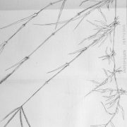
[Annotated ‘Ostitis[sic]’]

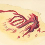
D 47 12RL On opening the muscles there was nothing to be seen but that the muscles were paler than usual & on opening the capsule of the joint the synovia was in excess & deeply died[sic] with blood, the synovial membrane was thicker than usual but not inflamed. The articular cartilage rather swollen and soft around the circumference of the humerus, but in no wise[?] inflamed or ulcerated, towards the ant: internal part of the articular surface of the scapular there was a slight abrasion, notched like of its circumference, it was smaller than a pea, but the cartilage was swollen & the bone was distinctly felt bare[?] in its centre, it was not inflamed or was there any sign of ulceration. The flexor Brachii[?] at its origin seemed injected & there was a bruise the size of a bean just below its tendon on the outside. The sub scapularis muscle had suffered, it presented the appearance of being severely bruised, ecchymosed through half its length and entire thickness. I could detect no rupture of its fibres, & its insertion was intact. The humerus & Scapular through half their length were congested, the periostium thickened and easily removed leaving the injected bone beneath. More particularly was this the case in certain parts such as near the insertion of the sub scapularis but the outside of the joint had shared in this apparently inflamed process but not to the same extent. There was osteitis of Humerus – see sketch

The history of the case of which the above is the post mortem is that on March 1st this horse collided during competition with a much heavier one, was struck on the near shoulder & knocked over. She fell on her side, but got up & walked to the infirmary with the leg dangling about like a broken one, thrown across the front of its fellow, the elbow being turned out, was placed in slings. When again examined next day when made to move the whole shoulder seemed to come out laterally the elbow turned out & the giving was so great that the animal leant entirely over to that side. I believe the 4th Condyle of humerus is knocked off & with it the attch of Portea Spinatus

March 6 – Blistered and consulted with Brown[?] & he thinks a fracture.

I divided this muscle in a dead limb and produced the deformity.

March 20 –  No better. Shoulder still out or seems to be every time he[sic] moves he can stand fair on limb, but can put no weight, can even make with, has a great knack of crossing his legs both front and rear Placed in slings and tied in. Pitch plaster &c. Placed the off fore on top of rear foot. Kicks the ground constantly. I think[?] from blister – but he does it day and night. Cause on P.M.E The lumbricoides

April 7 – Found with tetanus. Owing I think from chafes in slings.

[Annotated ‘Urine SpGz 1050. Ammonial & phosphates]

[[2]]
[Annotated ‘Lameness’]

Veterinary Regulations

Death Report

Regiment: 12 R Lancers

Station: Bangalore

Date: 8ber 1883

Troop or Battery: D

Number of Horses: 47

Sex: M

Age: 4

Disease: Tetanus

Date of Admission: April 7th 83

Date of Death: April 8th 83

Record of the Case

Copied from the Record Case Book

Case 62 D47

1883

7th April

Tetanus

This horse has been in the sick lines since March 1st with what I considered to be a fracture of the point of shoulders (near) there being extreme lameness & apparently dislocation. He was placed in slings. Lately he was found to be rather chafed round the chest as he was very uneasy and always kicking. This latter was not vice but was too continuous for such a long period not to attract attention

[[3]]

On visiting him this morning he was slightly blowing but feeding. On seeing him ¾ of an hour

later my suspicions were confirmed & I immediately administered, or rather tried to, a purgative. He lost nearly the whole dose. I then gave Carum[?] Indic: Ʒij with Heum Crosonis mxx in a ball. I managed to get this down. Removed to a loose box, & kept perfectly quite[sic], pulse normal, Temp 100o.

6.30pm Worse, the disease at its full height

At 9 am dropped down & died asphyxiated. [Annotated ‘8th’]

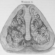
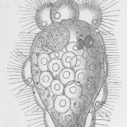
Autopsy. All viscera normal, but the stomach & duodenum contained I should say 500 Ascarides many of them over a foot in length, they nearly filled the duodenum

Remarks. Was it the chafe or Ascarides caused the tetanus. My view of the matter is that the chafe produced the mischief. The presence of this enormous number of parasites undoubtedly produced the great nuisance[?] which shewed itself in the continuous kicking there was no fracture of the shoulder, but ordeals[?] of humerus & severe bruising of Sub scapularis muscle

Discharged died

F Smith

[[4]]

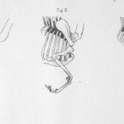
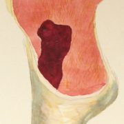
Osteitis of Humerus D 47

[Painting]

A section of the head of the humerus of D47. The medulla was exactly the above colour & the cancellous tissue was just as represented. I made no section of the scapular[sic] but I am sure it was the same.

[Transcription by Claudia Watts, KCL History, April 2019]

[FS/2/2/2/1/2]
[[1]]

N fore limb of I/3 horse injured at Kelbol[?]

The limb was an enormous size around the fetlock it was 17” in circumference & around the pastern 12”. There was the mark of an abscess on the outside and centre just above the fetlock. On removing the skin which was thick there was found enormous subcutaneous effusion of liquid and solid serum this was about 1 ½ “ to 2” in thickness. The superficial lymphatics were enlarged and contained coagulated lymph & a superficial abscess on front of fetlock After all this was cleaned away the Ext Pad just above the fetlock was directed downward and found to be slightly adherent to the bone & cut gently[?] the bone could also be felt nodular at this plane[.] On removing it on the joint the Capular opened was found to contain bloody muco-pus with air bubbles. The joint was laid completely open by dividing the later sesamoidal[?] lip and the synovial membrane was found scarlet and maroon extensive dislocation[?] of cartilage in several places varying from a about the size of sixpence to a pin point these were scarlet the cartilage in other places being partly absorbed the inflamed bone shown through black. There was extensive porcellaneous deposits in the joint no synovia. Partial dislocation of the met: found on the suffraginous sesamoid sheath the tendon was softened & adherent to each other & also to the fibro cartilage of sesamoids.

[Transcription by Claudia Watts, KCL History, April 2019]

Steel, John Henry – “A Manual of the Diseases of the Elephant and of his Management and Uses” (1885)

Terms of Use
This material has been identified as being free of known restrictions under copyright law, including all related and neighbouring rights, and is being made available under the Creative Commons, Public Domain Mark.